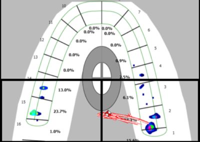
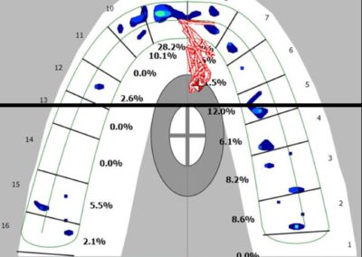
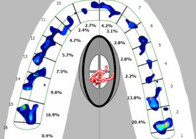
Force Distribution Patterns are diagnostic maps…
Over the life on an occlusion, the functional adaptation of the TMJ muscles, teeth and their supporting structures, airway, and neck posture can be physiologic, pathologic, or both. Whenever the stomatognathic system is engaged, it generates a repeating sequence of force with a traceable origin, direction, and intensity.
The mapping and recording of the distribution of these force measurements produce a unique, repeatable signature force pattern named the Habitual Force Pattern (HFP).
A HFP is a diagnostic map of the force measurements recorded while a patient swallows, squeezes, and taps firmly on a T-Scan recording sensor connected to a T-Scan® Occlusal Diagnostic System. Each HFP movie captures multiple sequential frames of force measurements displaying how the force distribution cycle is transferred on the occlusion. Force distribution cycles can be functional or dysfunctional.
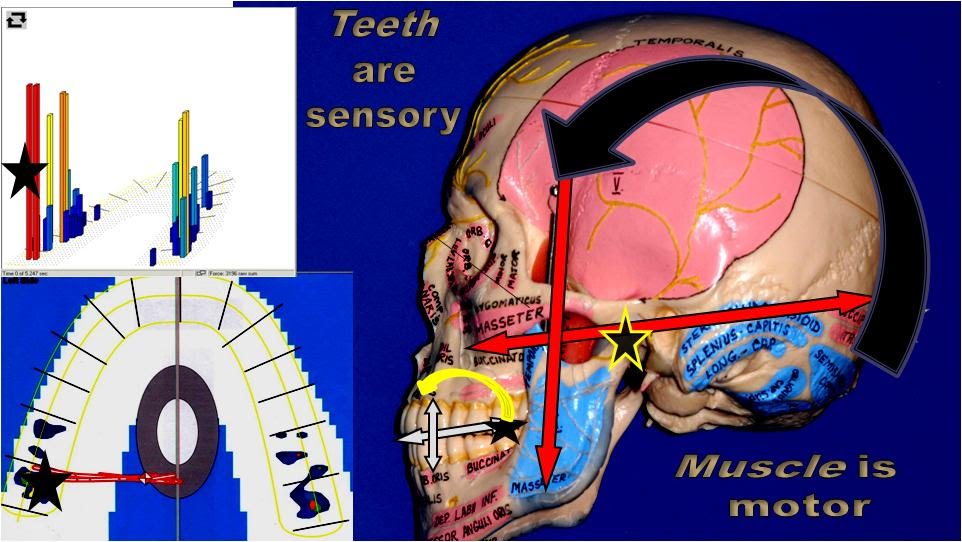
Clinical technique using the T-Scan® Occlusal Diagnostic System
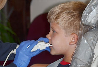
Recording a Habitual Force Movie
- Have the patient sit in the dental chair in a position similar to one adopted while eating and chewing.
- Insert the sensor in the patient’s mouth to start recording a 5- to 7-second movie.
- Ask the patient to swallow and then squeeze the sensor with clenched teeth.
- Ask the patient to tap firmly on the sensor three or four times and then to squeeze again with clenched teeth.
Recording a Skeletal Force Movie
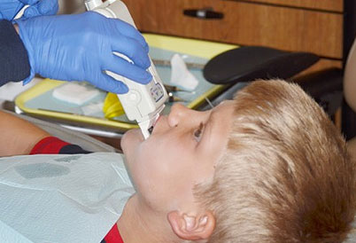
- Have the patient sit in a reclined position and apply a moderate amount of pressure to the front of the patient’s shoulders. This will pull the patient’s shoulders back, helping to immobilize the cervical muscles, the infrahyoid muscles, and the digastric muscles. This allows the joints and bones to follow their natural anatomy patterns.
- Insert the sensor in the patient’s mouth to start recording a 5- to 7-second movie.
- Ask the patient to swallow and then squeeze the sensor with clenched teeth.
- Ask the patient to tap firmly on the sensor three or four times and then to squeeze again with clenched teeth.
Recording a Centric Relation Force Movie
- Have the patient sit in a reclined position. Bimanually manipulate the patient’s jaw into centric relation.
- Have the chairside assistant insert the sensor in the patient’s mouth to start recording a 5- to 7-second movie.
- Still holding the patient’s jaw in CR, initiate closure into the sensor.

Recording a CR to CO Force Movie
- Have the patient sit in a reclined position. Bimanually manipulate the patient’s jaw into centric relation.
- Have the chairside assistant insert the sensor in the patient’s mouth to start recording a 5- to 7-second movie.
- Still holding the patient’s jaw in CR, ask the patient to swallow and squeeze the sensor while releasing your hold.
The figures below illustrate a single plane within each of the four force cycles. Images from the same plane are displayed in 2D on the left and in a 3D Contour format on the right.
The T-Scan® software allows practitioners to record, store, and assess the intensity and direction of the force exerted by mandibular natural teeth and/or prosthetics on the upper occlusal plane.
The digital articulating paper assigns a value to each tooth contact as patients tap on the recording sensor with minimal, maximal, then releasing force. These values can be viewed in either 2D or 3D.
The Center of Force marker is a compilation of all contacts by intensity at any given plane of the occlusal force cycle. While patients tap, the marker’s value is updated as the force exerted simulates a chewing cycle.
Each tap records a similar force sequence. As patients tap a second or a third time they tend to load the joints a little more with each tap. The final squeeze at the end of the recording represents the force on the occlusion when patients are in habitual interlock, also referred to as maximum intercuspal position (MIP). The recording of all force transfers throughout an occlusal cycle produces a unique, repeatable signature force distribution pattern named the Habitual Force Pattern (HFP). HFP patterns reveal how a bite will age, which teeth are involved in guiding and how the bite force is absorbed. Over the years, we have identified 6 patterns:


